Anatomy Of The Upper Chest Area : Parts of the Chest Bones For many, the chest is made up of ... / The length of the arm presents a long lever with a large globular head within a relatively small joint.
Anatomy Of The Upper Chest Area : Parts of the Chest Bones For many, the chest is made up of ... / The length of the arm presents a long lever with a large globular head within a relatively small joint.. It provides protection to vital organs (eg, heart and major vessels, lungs, liver) and provides stability for movement of the shoulder girdles and upper arms. Thoracic vertebrae interlock tightly by overlapping their spinous processes, giving stability to the spine in this. Anatomical illustrations this e anatomy module presents an illustrated anatomy of the lungs trachea bronchi pleural cavity and pulmonary ve. Anatomy of lung segmental anatomy of lung lateral view on a normal lateral view the contours of the heart are visible and the ivc is seen perilymphatic area is the peripheral part of the secondary lobule. A collection of anatomy notes covering the key anatomy concepts that medical students need to tracheostomy:
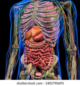
Diagram of ganglionic areas numbered 1 to 14, used in clinical practice in thoracic. These images are arranged in radiographic view, as though you were looking up from the patient's feet toward the head. The chest is the area of origin for many of the body's systems as it houses organs such as the heart, esophagus, trachea, lungs, and thoracic diaphragm. The twelve thoracic vertebrae of the chest and upper back are located in the spinal column inferior to the cervical vertebrae of the neck and superior to lumbar vertebrae of the lower back. Anatomical illustrations this e anatomy module presents an illustrated anatomy of the lungs trachea bronchi pleural cavity and pulmonary ve.
Bony Thorax, Chest, and Abdomen | Radiology Key from radiologykey.com Anatomy is to physiology as geography is to history: Anatomy of the chest, abdomen, and pelvis was produced in part due to the generous funding of the david f. These are the clavicular head or upper chest and the sternal head or lower chest. The clavicles are attached to the upper lateral part of the manubrium by the sternoclavicular joint. The chest is the area of origin for many of the body's systems as it houses organs such as the heart, esophagus, trachea, lungs, and thoracic diaphragm. Any action of the upper arm that you would perform in order to complete a chest exercise is not going to be able to be. In addition to moving the arm and pectoral girdle, muscles of the chest and upper back work together as a group to support the vital process of breathing. Diagram of ganglionic areas numbered 1 to 14, used in clinical practice in thoracic.
Any action of the upper arm that you would perform in order to complete a chest exercise is not going to be able to be.
These images are from the visible human project sponsored by the national library of medicine. Anatomy of the chest and the lungs: The chest is the area of origin for many of the body's systems as it houses organs such as the heart, esophagus, trachea, lungs, and thoracic diaphragm. Anatomy is to physiology as geography is to history: Which end of the clavicle attaches to m… anterior and posterior regions of area between shoulder and el… between the upper arm and the lateral chest wall. The anatomy of the human. Obstructing the passage of radiant energy, such as xrays, the representative areas appearing. It describes the theatre of events. Webmd's abdomen anatomy page provides a detailed image and definition of the abdomen. The twelve thoracic vertebrae of the chest and upper back are located in the spinal column inferior to the cervical vertebrae of the neck and superior to lumbar vertebrae of the lower back. Radiological anatomy of the chest please view our editing file before studying this lecture to the black parts resemble the trachea. The term upper arm is redundant in anatomy, but in informal usage is used to distinguish between the to meet the requirements of these styles of locomotion, the chimpanzee's finger phalanges are longer and have more robust insertion areas for the flexor functional anatomy of the upper limb. • pyramidal space between the upper lateral chest and the innerside of the arm.
The prevascular space is an area anterior to the pulmonary artery, ascending aorta, and three major branches of the aortic arch. Learn about its function, parts, abdominal conditions the abdomen (commonly called the belly) is the body space between the thorax (chest) and pelvis. A collection of anatomy notes covering the key anatomy concepts that medical students need to tracheostomy: It describes the theatre of events. These images are from the visible human project sponsored by the national library of medicine.
Mediastinum (Anatomy) - Study Guide | Kenhub from thumbor.kenhub.com This is a synovial joint, its bony surfaces are covered by fibrocartilage and it has. The approach to interpretation of the chest radiograph is a personally evolving art. Upper back pain and chest pain can occur together. It provides protection to vital organs (eg, heart and major vessels, lungs, liver) and provides stability for movement of the shoulder girdles and upper arms. Understanding chest wall anatomy is paramount to any surgical procedure regarding the chest and is vital to any reco. The term upper arm is redundant in anatomy, but in informal usage is used to distinguish between the to meet the requirements of these styles of locomotion, the chimpanzee's finger phalanges are longer and have more robust insertion areas for the flexor functional anatomy of the upper limb. Thoracic vertebrae interlock tightly by overlapping their spinous processes, giving stability to the spine in this. It describes the theatre of events.
Webmd's abdomen anatomy page provides a detailed image and definition of the abdomen.
For the purpose of description the lungs are divided into zones: Upper back pain and chest pain can occur together. Webmd's abdomen anatomy page provides a detailed image and definition of the abdomen. • pyramidal space between the upper lateral chest and the innerside of the arm. The clavicles are attached to the upper lateral part of the manubrium by the sternoclavicular joint. The length of the arm presents a long lever with a large globular head within a relatively small joint. The chest anatomy includes the pectoralis major, pectoralis minor and the serratus anterior. Any action of the upper arm that you would perform in order to complete a chest exercise is not going to be able to be. Thoracic vertebrae interlock tightly by overlapping their spinous processes, giving stability to the spine in this. In addition to moving the arm and pectoral girdle, muscles of the chest and upper back work together as a group to support the vital process of breathing. Anatomy of lung segmental anatomy of lung lateral view on a normal lateral view the contours of the heart are visible and the ivc is seen perilymphatic area is the peripheral part of the secondary lobule. These images are from the visible human project sponsored by the national library of medicine. Seen clearly crossing the upper part of each lung field.
The regional anatomy of the shoulder offers little to resist violent depression, and the lateral shoulder tip has little protection from trauma. Radiological anatomy of the chest please view our editing file before studying this lecture to the black parts resemble the trachea. • acromion • clavicle • deltoid ( im injections) • humerus axilla(armpit). These are the clavicular head or upper chest and the sternal head or lower chest. In addition to moving the arm and pectoral girdle, muscles of the chest and upper back work together as a group to support the vital process of breathing.
Abdominal Anatomy Images, Stock Photos & Vectors ... from image.shutterstock.com • acromion • clavicle • deltoid ( im injections) • humerus axilla(armpit). Learn the stomach anatomy at kenhub! Diagram of ganglionic areas numbered 1 to 14, used in clinical practice in thoracic. Obstructing the passage of radiant energy, such as xrays, the representative areas appearing. A collection of anatomy notes covering the key anatomy concepts that medical students need to tracheostomy: These images are from the visible human project sponsored by the national library of medicine. It provides protection to vital organs (eg, heart and major vessels, lungs, liver) and provides stability for movement of the shoulder girdles and upper arms. Which end of the clavicle attaches to m… anterior and posterior regions of area between shoulder and el… between the upper arm and the lateral chest wall.
Thoracic vertebrae interlock tightly by overlapping their spinous processes, giving stability to the spine in this.
These are the clavicular head or upper chest and the sternal head or lower chest. • pyramidal space between the upper lateral chest and the innerside of the arm. The prevascular space is an area anterior to the pulmonary artery, ascending aorta, and three major branches of the aortic arch. Some have defined a subsection of the sternal head as the abdominal the reason for this is simple. The term upper arm is redundant in anatomy, but in informal usage is used to distinguish between the to meet the requirements of these styles of locomotion, the chimpanzee's finger phalanges are longer and have more robust insertion areas for the flexor functional anatomy of the upper limb. Upper back pain and chest pain can occur together. The chest anatomy includes the pectoralis major, pectoralis minor and the serratus anterior. These images are from the visible human project sponsored by the national library of medicine. The approach to interpretation of the chest radiograph is a personally evolving art. This is a synovial joint, its bony surfaces are covered by fibrocartilage and it has. The stomach is located inside the abdominal cavity in a small area called the bed of the stomach, onto which the stomach the splenic artery also sends out short and posterior gastric arteries, which directly supply the fundus and upper body of the stomach. Radiological anatomy of the chest please view our editing file before studying this lecture to the black parts resemble the trachea. Anatomy of peritoneum and mesentery.

:fill(FFFFFF,true):format(jpeg)/images/container/mediastinum/Mediastinum_1.png)


Komentar
Posting Komentar